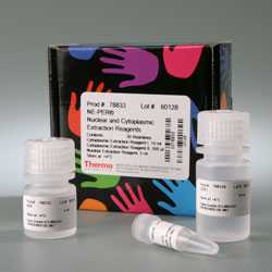
TheThermo Scientific NE-PER Nuclear and Cytoplasmic Extraction Kitprovides forefficient cell lysis and extraction of separate cytoplasmic and nuclear protein fractions in less than two hours. The NE-PER Kit is a nuclear protein extraction method that involves simple, stepwise lysis of cells and centrifugal isolation of nuclear and cytoplasmic protein fractions. A benchtop microcentrifuge, tubes and pipettors are the only tools required. The NE-PER Reagents efficiently solubilize and separate cytoplasmic and nuclear proteins into fractions with minimal cross-contamination or interference from genomic DNA and mRNA. Once desalted or diluted, the isolated proteins can be used to perform. immunoassays and protein interaction experiments, such as mobility shift assays (EMSA), co-immunoprecipitation (Co-IP) and pull-down assays.
Highlights:
- Fast– obtain nuclear and cytoplasmic fractions in less than two hours
- Proven– the NE-PER Reagent Kit is referenced in more than 950 distinct publications
- Scalable– two kit sizes for producing extracts from cells and tissues
- Convenient– simple instructions do not require ultracentrifugation over gradients
- Compatible– use for downstream assays, including Western blotting, gel-shift assays, protein assays, reporter gene assays and enzyme activity assays
Product Details:
There are a variety of methods available to isolate nuclei and prepare nuclear protein extracts. Most of the methods for preparing nuclear extracts are lengthy processes requiring mechanical homogenization, freeze/thaw cycles, extensive centrifugation or dialysis steps that may compromise the integrity of nuclear proteins. The NE-PER Nuclear and Cytoplasmic Extraction Reagent Kit is a reagent-based protocol that enables the stepwise lysis of cells, separation of the cytoplasm from the intact nuclease and then extraction of nuclear proteins away from genomic DNA and mRNA. This gentle process takes less than two hours and requires only a standard bench top centrifuge when using cultured cells. Furthermore, both active nuclear proteins and cytoplasmic proteins can be recovered from the same cell culture or tissue sample.
From two million cells, typical cytoplasmic protein yield is 200 to 500µg and typical nuclear protein yield is 100 to 200µg (at a concentration 1mg/mL). Typical cross-contamination between cytosolic and nuclear fractions is about 10%. The protein concentration of the nuclear extracts can be manipulated easily by varying the volume of nuclear extraction reagent (NER) used in the extraction without any significant loss in extraction efficiency. Specific extraction of nuclear proteins from cells is central to the success of many gene regulation studies. While a variety of methods exist to isolate nuclei and prepare nuclear protein extracts, most of these are lengthy processes requiring mechanical homogenization, freeze/thaw cycles, extensive centrifugation or dialysis steps that may compromise the integrity of many fragile nuclear proteins. The NE-PER Kit overcomes these problems, providing high-quality, concentrated nuclear protein extracts for use in EMSA and other analysis techniques.
 |
| Total protein profile of cytoplasmic and nuclear extracts prepared from a variety of mammalian cell lines using NE-PER Reagents.Protein was quantitated using Pierce Micro BCA Protein Assay Reagent (Part No. 23235). Values are the average of two separate isolations. |
 |
| Western blots of specific proteins fractionated using NE-PER into cytosolic extracts (C) and nuclear extracts (N).Two million A549 cells were lysed using Pierce Nuclear and Cytoplasmic Extraction Reagent Kit (NE-PER). Samples were normalized for protein concentration using Pierce BCA Protein Assay. 10µg of each cytosolic and nuclear extract sample was analyzed by 4-20% SDS-PAGE and Western blotted using specific antibodies diluted 1:1000 (HSP90, Ran, MEK1, MSH2, MCM2, HDAC1, DDX3, TopoIIb, NFκB) or 1:10000 (p53, GSK3β, β-catenin, GAPDH). Anti-mouse (H+L) HRP or anti-rabbit (H+L) HRP diluted 1:25,000 was used as the secondary antibody with SuperSignal West Dura Chemiluminescent Substrate for detection. |
 |
| Total protein profile of cytoplasmic and nuclear extracts prepared from different mouse tissues.Swiss Webster mouse tissues (40mg) were harvested, rinsed with PBS and lysed using the NE-PER Nuclear and Cytoplasmic Extraction Reagent Kit. Extracts were quantified using the Pierce 660nm Protein Assay Reagent (Part No. 22660). Values are the average of two separate isolations. |
 |
| Western blots of specific proteins from fractionated tissues.Cytoplasmic and nuclear extract (10µg each) from different mouse tissue fractionated using the NE-PER Nuclear and Cytoplasmic Extraction Reagent Kit was analyzed by 4-20% SDS-PAGE and Western blotting. Primary antibodies specific for the target proteins were diluted 1:1,000 (SP1, HDAC2 and NFκB p65, or 1:10,000 (GAPDH). Anti-Rabbit (H+L) HRP (Part No. 31460) diluted 1:25,000 was the secondary antibody and SuperSignal West Dura Chemiluminescent Substrate (Part No. 34076) was used for signal detection. |
 |
| Chemiluminescent EMSA of four different DNA-protein complexes.DNA binding reactions were performed using 20fmol biotin-labeled DNA duplex (1 biotin per strand) and 2µL (6.8µg total protein) NE-PER Nuclear Extract prepared from HeLa cells. For reactions containing specific competitor DNA, a 200-fold molar excess of unlabeled specific duplex was used. |
Product Reference:
- Nien-Pei, Tsai,et al.(2010). Dual action of epidermal growth factor: extracellular signal-stimulated nuclear-cytoplasmic export and coordinated translation of selected messenger RNA.J. Cell Biol.188 (3), 325-333.
References:
- Trotter, K. W. and Archer, T. K. (2004). Reconstitution of glucocorticoid receptor-dependent transcriptionin vivo.Molecular and Cellular Biology 24(8), 3347-3358.
- Liu, H.B.,et al. (2003). Estrogen receptor alpha mediates estrogen’s immune protection in autoimmune disease.J. Immunol. 171(12), 6936-6940.
- Yamada, N. A.et al. (2003). Variation in the extent of microsatellite instability in human cell lines with defects in different mismatch repair genes.Mutagenesis 18(3), 277-282.
- Kim, K.et al. (2002). Expression of a functional Drosophila melanogaster N-acetylneuraminic acid (Neu5Ac) phosphate synthase gene: evidence for endogenous sialic acid biosynthetic ability in insects.Glycobiology 12(2), 73-83.
- Lee, S. and River, C. Role played by hypothalamic nuclear factor-κB in alcohol-mediated activation of the rat hypothalamic-pituitary-adrenal axis.Endocrinology 146(4), 2006-2014.
- Penheiter, S. G.et al. (2002). Internalization-dependent and -independent requirements for transforming growth factor β receptor signaling via the Smad pathway.Molecular and Cellular Biology 22(13), 4750-4759.
- Shih, H-P.et al. (2002). Identification of septin-interacting proteins and characterization of the Smt3/SUMO-conjugation system in Drosophila.Journal of Cell Science 115(6), 1259-1271.
- Yamada, N. A. and Farber, R. A. (2002). Induction of a low level of microsatellite instability by overexpression of DNA polymerase β1.Cancer Research 62, 6061-6064.
- Adilakshmi, T. and Laine, R.O. (2002). Ribosomal protein S25 mRNA partners with MTF-1 and La to provide a p53-mediated mechanism for survival or death.J. Biol. Chem. 277(6), 4147-4151.
- Sanceau, J. (2002). Interferons inhibit tumor necrosis factor-a-mediated matrix metalloproteinase-9 activation via interferon regulatory factor-1 binding competition for NF-κB.J. Biol. Chem. 277, 35766-35775.
- Smirnova, I. V.et al. (2000). Zinc and cadmium can promote rapid nuclear translocation of metal response element-binding transcription factor-1.Journal of Biological Chemistry 275(13), 9377-9384.
- Current Protocols in Molecular Biology (1993).
- Zerivitz, K. and Akusjarvi, G. (1989).Gene Anal. Tech. 6, 101-109.
- Dignam, J.D.,et al. (1983).Nucleic Acids Res. 11, 1475-1489.
|


